Think about sorting a basket of fruits. Before you decide what to do with them, you first separate apples from oranges, ripe fruits from unripe ones, and damaged fruits from fresh ones. That simple act of sorting helps you make better decisions.
Microbiology works in a similar manner. Before researchers get a chance to identify a microbe in detail, they often want a quick way to have a look at what they are dealing with. One of the most widely used methods for this is gram staining.
Despite the availability of modern equipment and molecular techniques in the laboratories, Gram Staining is still valuable for microbiological investigation. It provides useful information in a short time and can guide subsequent identification and testing.

Gram staining is a laboratory technique that is used to differentiate bacteria into two groups, Gram-positive or Gram-negative, based on the composition of their cell walls. When bacteria are treated with various staining agents, they respond differently and have distinct colours when observed under the microscope. This easy separation is useful for microbiologists, healthcare professionals, researchers, and students. In many cases, gram staining is the first observation when studying bacteria.
Just as a map helps travellers understand where they are before continuing their journey, gram staining provides an initial clue to what a lab may be dealing with before it proceeds to other more specific tests.
Every bacterial cell is built differently. Some have thicker cell walls and others have thinner layers with extra outer components. The principle of gram staining is based on these differences of structures.
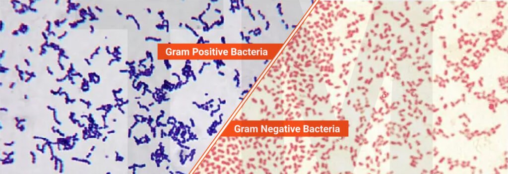
One key factor is the peptidoglycan layer in the bacteria cell wall. In Gram-positive bacteria this layer is relatively thick, and they are able to retain the primary stain in the staining procedure and will look purple or violet when observed under the microscope. The peptidoglycan layer of Gram-negative bacteria is very thin and covered by an outer membrane, which makes it impossible to stain them the same way. Due to this structure, they do not retain the primary stain but instead take up the counterstain and turn pink or red.
By observing these colour differences, microbiologists can easily identify the Gram-positive and Gram-negative bacteria. The visual differentiation of microorganisms is what makes this technique useful in laboratory analysis and microbiology learning.
The application of the gram staining is more than classroom learning. In clinical laboratories, it helps in the preliminary evaluation of patient specimens. It is used to track the contamination of microorganisms in the food and beverage industry. It is used in research laboratories to investigate bacterial cultures and to verify the purity of microbial isolates.
Environmental microbiologists also use this method to investigate microorganisms from soil, water, and other environmental samples. Its adaptability makes it suitable for use in many fields of microbiological work. As results can be generated rapidly, it is generally used as a screening test prior to more specific testing.
The significance of gram staining extends beyond simply classifying bacteria. It assists laboratories in:
Since the method is relatively simple and inexpensive, it remains accessible to laboratories with different levels of resources.
To numerous students stepping into a microbiology laboratory for the first time, microscopic organisms can seem difficult to understand. Textbooks explain the structures of bacteria, but looking at those differences yourself under a microscope is a much more powerful way to learn. This is one reason gram staining continues to be the cornerstone activity in microbiology education. Students benefit because it
The principle of gram staining is usually one of the first concepts students learn when studying bacterial identification. By performing the procedure themselves, they gained a better idea of how microbiologists study and classify microorganisms.
However gram staining is a useful technique; it also has a few limitations. Not all microorganisms have a clear reaction to the staining process. Certain bacteria are variably stained, while some require further testing prior to identification.
The result may also be influenced by the quality of the specimen. Interpretation may be affected by improper smear preparation, damaged cells, or contaminants. Therefore, staining observations are usually supplemented by culture, biochemical, or molecular analysis to obtain a complete understanding of the microorganism.
Microbiology depends on methods that yield dependable information with a practical output in an efficient manner. Gram staining continues to serve this purpose by offering a simple way to visualize bacterial characteristics that may be used to guide additional laboratory analyses.
Whether it is to help students to learn basic principles of microbiology or professionals to carry out routine day-to-day laboratory work, the usefulness is always clear. Although numerous modern techniques are now available, gram staining remains an important tool in microbiology, providing valuable information by a simple and accessible technique.
A. Gram staining offers a fast visual result of the bacteria, whereas culture methods are based on the growth of microorganisms to achieve advanced identification and analysis.
A. No, it can be performed using normal equipment found in any microbiology lab.
A. Yes.
A. To classify bacteria and guide further laboratory investigations.
A. Common errors include applying stains for incorrect durations, over-decolorization, under-decolorization, preparing thick smears, or using damaged bacterial cultures. These factors can influence the final appearance of the cells.
At some point all microbiologists have experienced this situation at least once. A clinical specimen arrives at the laboratory. It...
Read More
Most of us have come across algae at some point floating on the surface of a pond, growing along riverbanks,...
Read More
In their daily life, a microbiologist works with a variety of samples. And the one who works with Stool Samples...
Read More
In microbiology laboratories, collecting a specimen is only the first step in obtaining accurate results. The state of the sample...
Read More
In every microbiology laboratory, careful handling of microorganisms is a key priority. Even a minor error in the transfer of...
Read More
How can I help you today?