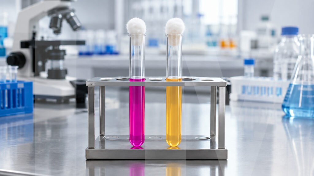
A small tube sits on a microbiologist’s bench, looking almost ordinary. Its orange medium waits for one biological event to reveal itself.
After inoculation, the medium may begin to change. A shift toward pink can turn a quiet observation into a useful biochemical clue.
That colour change reflects urease activity. The enzyme breaks down urea and releases products that increase the medium’s alkalinity.
This simple reaction is the basis of the urease test. It enables microbiologists to examine organisms by their ability to produce urease.
The urease test detects whether a microorganism can produce the enzyme urease. Urease acts on urea as its substrate.
Urea hydrolysis yields ammonia. The produced ammonia increases the pH of the surrounding medium and makes it alkaline. This alkaline shift causes the medium to change from orange toward pink.
Urea Agar Base is a suitable medium for the observation of this reaction. It is especially good for detecting Proteus vulgaris, Micrococci and paracolon organisms. It also differentiates between organisms with different urease reactions.
Christensen developed a modified formulation to detect rapidly urease-positive Proteus organisms and delayed reactions in other Enterobacteriaceae.
The composition of Urea Agar Base combines nutrients, salts, an energy source, a solidifying agent, and a pH indicator. Each component supports a specific function within the medium.
| Ingredients | Gms / Ltr |
| Peptone | 1.000 |
| Agar | 15.000 |
| Phenol red | 0.012 |
| Sodium chloride | 5.000 |
| Dextrose (Glucose) | 1.000 |
| Disodium phosphate | 1.200 |
| Monopotassium phosphate | 0.800 |
Peptone is a source of nutrients for the growth of microbes. Agar is the solid matrix required to make the medium as a slant.
Phenol red was employed as a pH indicator. Sodium chloride is used to maintain the osmotic balance of the preparation.
Dextrose is an energy source for organisms. The buffering system is a mixture of monopotassium phosphate and disodium phosphate.
Together, these components form an environment in which urease-associated changes in the medium can be detected.
The principle of the urease test is based on the reaction of urease with urea. If an organism produces urease, the enzyme hydrolyses urea during incubation.
This reaction also liberates ammonia and increases the alkalinity. Phenol red then responds to the resulting pH change.
In Urea Agar, the medium appears orange before the reaction occurs. A positive reaction changes the colour from orange to pink as alkalinity develops.
There are a few components that facilitate this reaction without performing the same role. Peptone supplies essential nutrients, and dextrose is a source of energy.
Sodium chloride is important in maintaining osmotic balance. Phosphates act as buffer substances in the medium.
The formulation also uses reduced peptone and a reduced buffering system. These changes help the medium detect smaller amounts of alkali.
Adding glucose also forms part of Christensen’s modification. These changes help reveal delayed urease reactions.
A colour change alone does not always prove urease activity. Other processes can also increase alkalinity during prolonged incubation.
Protein hydrolysis can raise the pH as it produces alkaline products. Hence, excess amino acids can also result in a false-positive reaction.
A medium without urea can serve as a negative control. This control helps identify alkaline changes unrelated to urea hydrolysis.
The test also has an important analytical limitation. Urea test media rely on alkalinity formation, so they cannot determine the absolute rate of urease activity.
For this reason, microbiologists should interpret the reaction within the complete identification process. The result may give valuable evidence without serving as the sole basis of identification.
Urease detection is used by microbiologists to differentiate between the organisms that can hydrolyse urea. This biochemical feature can support identification workflows.
Proteus vulgaris represents an important organism associated with the intended use of Urea Agar Base from TM Media. Micrococci and paracolon organisms also appear within its stated application.
The reaction can also help distinguish rapid urease-positive organisms from organisms showing delayed urease activity. This distinction becomes useful when several organisms produce different biochemical profiles.
A urea hydrolysis test therefore connects enzyme activity with an observable laboratory result. The visible reaction can support organism identification.
However, the term ‘specific urease test’ needs careful interpretation. The test detects urease-related alkalinity, but it does not measure the absolute enzyme rate.
The method employs a defined substrate, nutrients, controlled preparation and a pH indicator.
A simple colour change can carry meaningful information when the chemistry behind it is understood. Urea Agar Base turns urease activity into a visible biochemical reaction.
The process begins with urea and ends with an alkaline reaction.
Controls are needed, as unrelated alkaline reactions could give false positive results. Proper timing remains essential during interpretation.
The test shows how microbial activity can be turned into laboratory evidence, from a silent orange slant to an observable pink reaction.
Want to explore the Urea Agar Base in more detail? (Link here)
A. Urease is commonly produced by Proteus vulgaris. Other urease producers may also give positive reactions, especially when conditions are suitable for testing.
A. The slant gives a firm surface for inoculation. The prepared medium is in a suitable form for observation of the reaction.
A. Protein hydrolysis and prolonged incubation can give false positive reactions.
A. Urease acts on urea and breaks it down during the reaction.
A. No. This test is designed for the alkaline reaction which is associated with the hydrolysis of urea. It does not control the rate of urease activity.
A microbiologist studies a bacterial culture that looks no different from many others. Yet, its metabolic capabilities may be an...
Read More
A microbiologist often looks beyond what appears on a culture plate. A tiny change in colour can reveal an important...
Read More
A colony appearing on an agar plate may look like a simple result. Yet, that small colony reflects everything the...
Read More
A microbiologist receives a mixed sample and prepares a fresh culture plate. The next question seems simple: which organism will...
Read More
A microbiologist opens two Petri plates after incubation. Both plates contain the same sample. One plate shows clear, well-separated colonies....
Read More
How can I help you today?
