
If one imagines the deadliest microbial infection that affects the weakest and the most sensitive fraction of our population, one could hear the loud echoing voices of pneumonia inside their heads. Pneumonia got recent worldwide attention after researchers discovered that it was the leading consequence and cause of death amongst individuals infected with COVID-19. But a closer study reveals that COVID-19 is far from playing the protagonist when it comes to the deadly play of pneumonia. Actually, Streptococcus pneumoniae (S.pneumoniae) is the leading cause of pneumonia infection which claims millions of lives every year.
Streptococcus Pneumoniae is a gram positive, lancet shaped bacterium which is fastidious in nature and targets the lower respiratory tract of humans. Although today we have many biochemical solutions like vaccine and antibiotics for curing the infected individuals, still a large percentage of infants below five years of age and elders above 70 years of age become victims of this horrific bacteria. There is no turning away from the fact that this gram-positive bacterium could be titled as the “Killer of Youth”.
The biggest killer in India
In 2018, India witnessed the second highest number of deaths of children under the age of five due to pneumonia. Over 1,27,000 young Indian infants fell prey to this deadly lower respiratory tract infection that year. The bacteria causes the air sacs to be filled with puss and toxic biochemical which suffocate the patients to death. Bronchial Pneumonia caused by Streptococcus Pneumoniae is highly contagious in nature, therefore it spreads easily at community level resulting in increased cases of community acquired pneumonia. Fever, cough, sputum production, rigors, pleuritic chest pain, dyspnea, tachycardia, bronchial breath sounds and crackles on auscultation are some of the symptoms of lower respiratory tract pneumonia.
The attack mechanism
S.pneumoniae has a very mechanistic strategy of capturing the lung surface for rapid proliferation of the infection. During infection, mesothelium layer comprising of mesothelial cells acts as the first line of defense against the deadly pathogens to prevent the foreign colonization of pleura. Mesothelial cells can release a large range of pro-inflammatory cytokines which subsequently recruit immune cells at the pleural cavity to fight against the pathogens.
But years of bacterial evolution have given a special ability to Streptococcus Pneumoniae which claims victory even before the war is drawn upon by mesothelial cells against them. Streptococcus Pneumoniae bacterial cells secrete a cytotoxic protein called pneumolysin which acts like bullets to form large pores in membranes of mesothelial cells. This leads to lysis of mesothelial cells and a free passage of pathogens inside the pleural cavity where bacterial cells proliferate and start attacking lung surface, bronchi and other vital parts of human lung. Such infections usually lead to dangerous Empyema, a condition where pus or bacterial culture gets accumulated in the pleural cavity.
The recent cause of concern
To add to the degree of horridness of Streptococcus Pneumoniae infection, recent studies show that it has become resistant to many antibiotics and therefore treating bacterial pneumonia has become one of the challenges. When the youngest of our society become endangered to infection, so does the future of the society. Hence, it is highly important to treat our younger ones and other infected individuals who contribute towards the society.
The Diagnosis
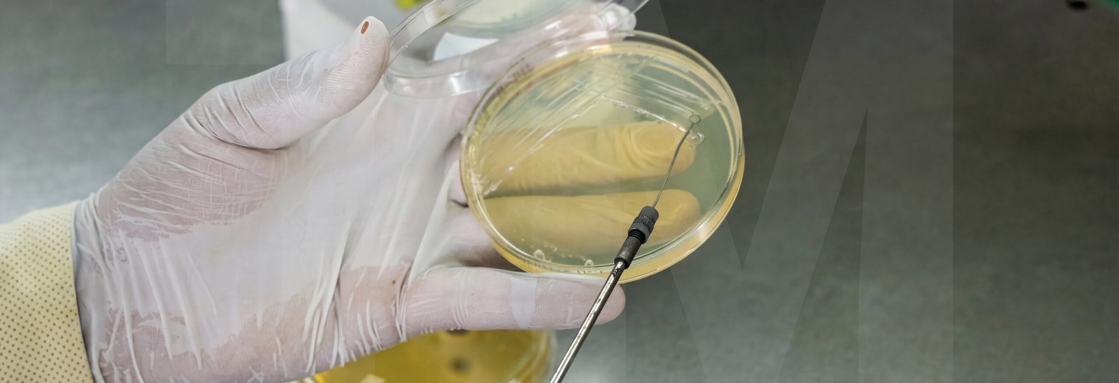
Since many years, pneumonia caused by Streptococcus Pneumoniae is diagnosed on sheep blood agar plates. The agar media used in such plates is rich in red blood cells which are destroyed/lysed by bacterial toxins. S.pneumoniae produces hydrogen peroxide which causes alpha hemolysis or “green” hemolysis by oxidizing hemoglobin to green methemoglobin. This forms a green zone around Streptococcus Pneumoniae colonies formed on the sheep blood agar plate.
Unfortunately, a group of Streptococcus viridans also exhibit alpha hemolytic activity. Thus, this method creates a lot of confusion and false results as both types of colonies are similar.

Since evolution has made Streptococcus Pneumoniae resistant to a group of antibiotics, researchers have exploited this fact to create a unique strategy for selectively diagnosing S.pneumoniae colonies. Adding 5ug of gentamycin to 5% sheep blood agar increases the yield of Streptococcus Pneumoniae from respiratory secretions and apparently suppresses the growth of other bacteria like S.viridans.
TM Media offers such a customized product (TMP 034), which allows the technicians to selectively cultivate or diagnose S.pneumoniae on Sheep Blood Agar plates infused with Gentamycin (5ug/plate). These ready to use culture media plates save the professionals from spending a great deal of time in weighing, measuring, autoclaving, pouring, etc.
A microbiologist finishes inoculating a culture plate and places it inside the incubator. The next day, the plate is covered...
Read More
Every microbiologist has seen this happen. A clinical specimen from a patient with suspected bacterial meningitis is received. The sample...
Read More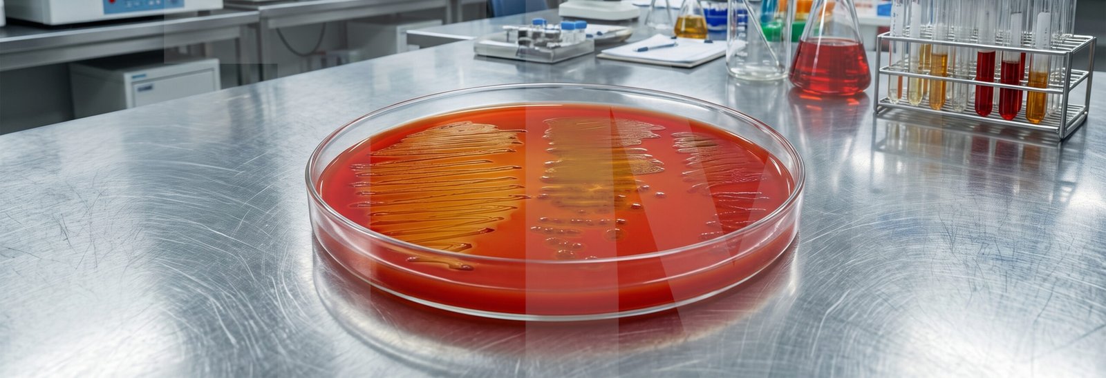
At some point all microbiologists have experienced this situation at least once. A clinical specimen arrives at the laboratory. It...
Read More
Most of us have come across algae at some point floating on the surface of a pond, growing along riverbanks,...
Read More
In their daily life, a microbiologist works with a variety of samples. And the one who works with Stool Samples...
Read More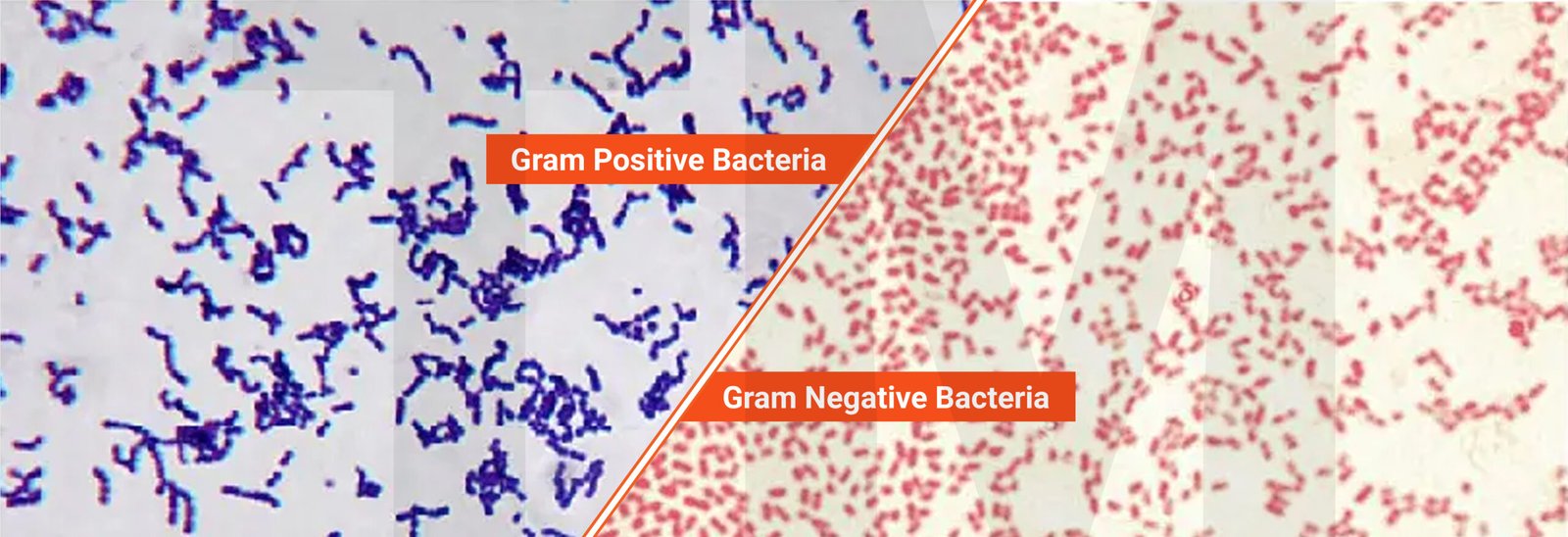
Think about sorting a basket of fruits. Before you decide what to do with them, you first separate apples from...
Read More
How can I help you today?